
In the dynamic landscape of modern education and professional development, particularly within the demanding fields of medicine and healthcare, tools that can simplify complex subjects while maintaining absolute accuracy are invaluable. The Human Anatomy Atlas 2018: Complete 3D Human Body stands out as a pioneering educational application, meticulously crafted to offer an immersive, detailed, and biologically accurate exploration of the human form. Designed for a broad audience, from budding medical students grappling with the intricacies of human physiology to seasoned professionals seeking to refresh their knowledge or explain conditions to patients, this application transcends traditional learning methods by providing an unparalleled virtual human body experience.
At its core, Human Anatomy Atlas 2018 is more than just an app; it is a comprehensive digital atlas that brings anatomy to life in breathtaking three dimensions. Eschewing the limitations of two-dimensional diagrams and static models, it ushers users into an interactive world where the human body can be deconstructed, examined, and understood with unprecedented depth. This innovative approach to anatomical study is not merely a technological advancement but a pedagogical revolution, enabling a more intuitive and engaging learning process that caters to diverse learning styles and professional requirements. Endorsed by medical experts, the meticulously rendered male and female 3D models within the atlas are a testament to its commitment to biological fidelity, ensuring that every bone, muscle, artery, and organ is depicted with scientific precision. This dedication to accuracy makes it a trusted resource in an arena where even the slightest misrepresentation can have significant implications.
Unveiling the Human Form in Three Dimensions: A Journey of Discovery
The cornerstone of Human Anatomy Atlas 2018’s utility lies in its extraordinary 3D visualization capabilities. It transforms the often-challenging task of understanding spatial relationships within the human body into an intuitive and interactive experience. Users are not passive observers but active participants in their learning journey, capable of manipulating, dissecting, and analyzing anatomical structures with a level of detail previously unimaginable outside of cadaver labs. The application provides a complete 3D reference of both male and female human bodies, allowing for a comparative study that highlights the nuances and distinctions between the sexes, which is crucial for a holistic understanding of human biology.
Anatomical Precision and Interactive Dissection
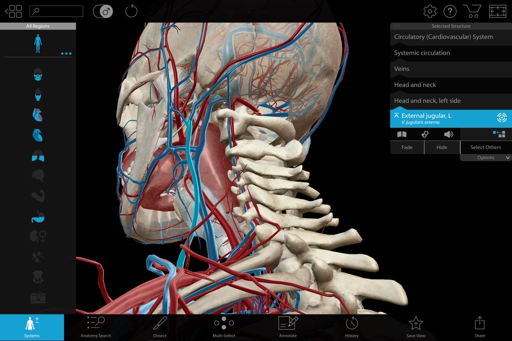
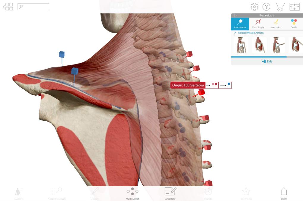
The commitment to anatomical accuracy is perhaps the most lauded feature of Human Anatomy Atlas 2018. The 3D models are not generic representations but are biologically accurate, validated by a panel of medical experts to ensure that every structure, from the smallest ligament to the largest organ system, adheres to scientific standards. This level of verification provides a strong foundation of trust for users who rely on the atlas for critical learning and reference. The interactive nature of the models allows for “layer-by-layer” examination, mimicking the process of a physical dissection. Users can systematically peel back layers of skin, muscle, and tissue to reveal the underlying structures. Imagine being able to isolate and inspect a single bone, trace the path of an artery through a complex network of tissues, or observe the intricate interplay of muscles during movement—all within a virtual environment. This capability is particularly beneficial for visual learners, who can better grasp the three-dimensional organization of the body when they can rotate, zoom, and manipulate structures at will. The ability to examine cross-sections of bones, muscles, and arteries further enhances this understanding, offering insights into the internal architecture and relationships between different tissues. Furthermore, the inclusion of animations depicting various body parts in action brings a dynamic dimension to the learning process, illustrating physiological functions such as muscle contractions, joint movements, and organ system operations in a clear and engaging manner. These animated sequences are crucial for understanding not just what structures are present, but how they function together to sustain life.
From Gross Anatomy to Microscopic Wonders
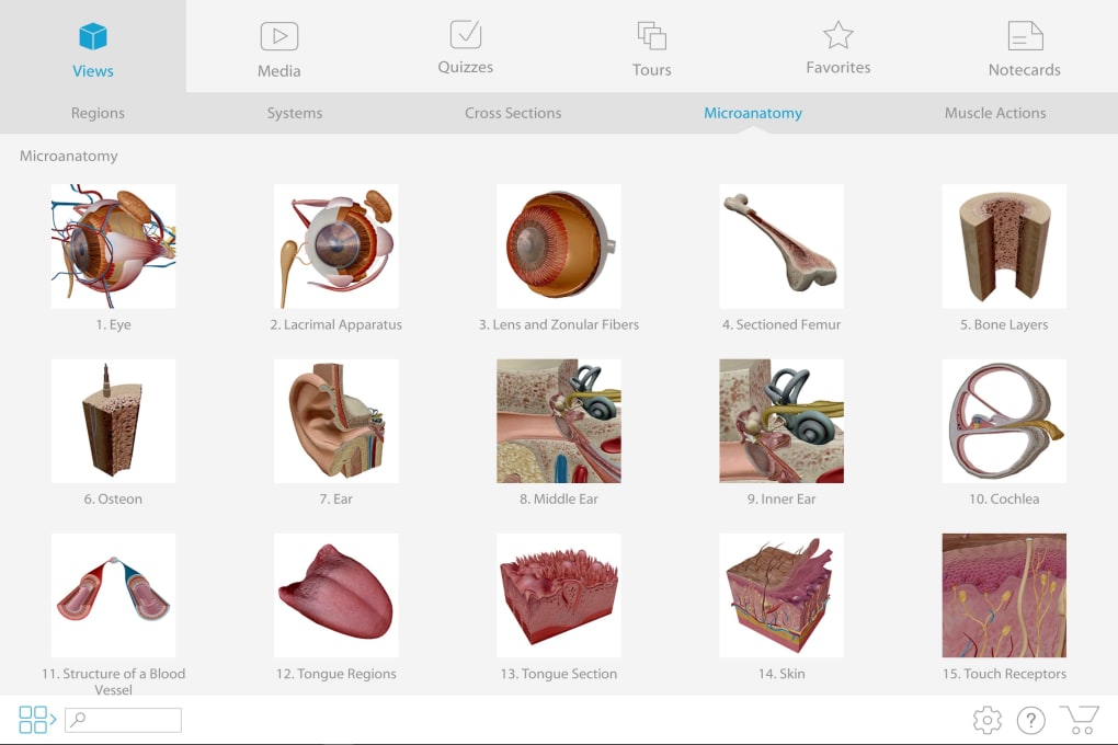
While many anatomical atlases excel at presenting gross anatomy, Human Anatomy Atlas 2018 distinguishes itself by seamlessly bridging the gap between macroscopic and microscopic perspectives. The application allows users to zoom far beyond what is typically visible to the naked eye, delving into the realm of microanatomy. This feature is particularly groundbreaking, as it enables students and professionals to explore the minute structures that are fundamental to understanding physiological processes at a cellular and tissue level. For instance, one can zoom into the level of microanatomy to look at nephrons within the kidney, the intricate filtering units responsible for urine formation. Similarly, users can explore alveoli in the lungs, the tiny air sacs where gas exchange occurs, or the elaborate structures of a neuron in the brain. This capability is vital for comprehending how macroscopic organ functions are underpinned by microscopic cellular and tissue arrangements. The transition from a full-body view to these minuscule components is smooth and intuitive, providing a comprehensive and integrated understanding of the human body’s hierarchy of organization. This holistic approach ensures that learners appreciate the interconnectedness of all anatomical levels, from the grand architecture of organ systems down to the cellular machinery that drives life. Such detailed exploration at the micro level is not only fascinating but essential for disciplines like histology, pathology, and advanced physiology, making Human Anatomy Atlas 2018 an indispensable resource for a wide array of medical and scientific studies.
A Comprehensive Learning Companion: Beyond Visuals
While the stunning 3D visuals are a primary draw, Human Anatomy Atlas 2018 recognizes that a truly comprehensive educational tool must offer more than just pictures. It integrates a wealth of textual information and interactive assessment features to complement the visual learning experience, creating a multifaceted resource that caters to various learning preferences and reinforces knowledge effectively. This dual approach ensures that users not only see anatomical structures but also understand their names, functions, clinical significance, and relationships to other parts of the body.
Rich Textual Resources and Multilingual Support
Accompanying the highly detailed 3D models is an extensive digital encyclopedia of the human body. This encyclopedic content provides in-depth textual descriptions for every anatomical structure presented visually. For each bone, muscle, nerve, vessel, and organ, users can access detailed explanations covering its nomenclature, origin, insertion, innervation, blood supply, actions, and clinical correlations. This integration of visual and textual information is crucial for building a robust understanding of anatomy, moving beyond simple identification to functional comprehension. The textual material is meticulously researched and peer-reviewed, ensuring its scientific accuracy and relevance to medical curricula. What further enhances the accessibility and global utility of Human Anatomy Atlas 2018 is its multilingual support. The digital encyclopedia, along with the application’s interface, is available in multiple languages. This feature is immensely beneficial for international students and professionals, allowing them to learn and reference anatomical terms in their native language or a preferred academic language, thereby breaking down linguistic barriers to learning and making the complex subject matter more approachable for a wider audience. The ability to switch between languages also serves as an excellent tool for language learners in the medical field, helping them to master anatomical terminology across different linguistic contexts. The sheer volume and quality of this integrated textual content elevate the atlas from a mere visualization tool to a powerful study and reference companion. It enables users to delve deeper into the context of each anatomical part, understanding its role within the larger system and its clinical significance, which is paramount for practical application in healthcare.
Interactive Quizzes for Knowledge Reinforcement
Learning anatomy effectively requires not just absorption of information but also active recall and self-assessment. Human Anatomy Atlas 2018 incorporates a robust system of anatomy quizzes designed to test and reinforce users’ knowledge in an engaging and effective manner. These quizzes are not generic multiple-choice questions but are intelligently designed to challenge users on their understanding of anatomical identification, spatial relationships, and functional knowledge. Users can test their knowledge by taking part in various types of quizzes, which may include identifying structures based on their 3D models, answering questions about their functions, or locating specific parts within a complex anatomical region. The immediate feedback provided by the quizzes allows users to identify areas where their understanding is strong and pinpoint areas that require further study. This iterative process of learning, testing, and reviewing is a proven pedagogical method for improving retention and mastery of complex subjects. For medical students preparing for exams, these quizzes offer an invaluable self-assessment tool, helping them to gauge their preparedness and focus their study efforts efficiently. For professionals, the quizzes serve as a convenient way to refresh and solidify their anatomical knowledge, ensuring that their foundational understanding remains sharp and current. The interactive and gamified nature of these assessments can also make the learning process more enjoyable and less intimidating, encouraging consistent engagement with the material. By combining detailed visuals and rich textual information with practical, interactive assessments, Human Anatomy Atlas 2018 creates a complete learning ecosystem, empowering users to not only explore but also truly master the intricacies of human anatomy. Whether one is a medical student striving for academic excellence or a professional committed to continuous learning, the blend of visual exploration, comprehensive information, and self-assessment tools makes this app an exceptionally useful and effective resource.
Designed for Diverse Learners and Professionals: Bridging Gaps in Medical Education
The versatility of Human Anatomy Atlas 2018 makes it an indispensable asset across the spectrum of medical and healthcare education and practice. Its intuitive design and comprehensive content are tailored to meet the distinct needs of various user groups, from those just beginning their anatomical journey to those who apply this knowledge daily in clinical settings. The app’s ability to simplify complex concepts without sacrificing detail makes it a bridge over many of the common challenges faced in learning and teaching human anatomy.
Empowering Medical Students
For medical students, the study of human anatomy is often considered one of the most challenging yet foundational aspects of their curriculum. Traditional methods involving textbooks, 2D diagrams, and cadaver dissection, while essential, can sometimes be overwhelming. Human Anatomy Atlas 2018 provides a critical supplementary tool that transforms this challenging subject into an accessible and interactive learning experience. Students can use the app to visualize structures that might be difficult to observe or fully comprehend in a dissection lab, especially if access to cadavers is limited or the structures are particularly intricate. The ability to rotate, zoom, and layer anatomical models helps in understanding the spatial relationships between different body parts, a concept notoriously difficult to grasp from flat images. This virtual exploration allows students to prepare for lab sessions, review after lectures, and solidify their understanding for exams. For instance, prior to a dissection, a student can virtually “dissect” the region in the app, familiarizing themselves with the expected structures and their arrangements. Post-lab, they can review what they observed, comparing it with the perfectly rendered models. The detailed textual information, coupled with interactive quizzes, further reinforces their learning, helping them to memorize names, functions, and clinical correlations. The app can also be a lifeline for students who struggle with traditional memorization techniques, as the visual and interactive nature promotes a deeper, more conceptual understanding. By providing a personalized, on-demand anatomical reference, Human Anatomy Atlas 2018 empowers medical students to take control of their learning, leading to improved comprehension, better retention, and ultimately, greater success in their studies.
A Vital Tool for Healthcare Professionals
Beyond the academic realm, Human Anatomy Atlas 2018 offers significant utility for a wide array of healthcare professionals. Doctors, nurses, physical therapists, chiropractors, and other allied health professionals can leverage the app as a quick and reliable reference tool in their daily practice. For clinicians, the app can serve as an invaluable resource for patient education. Explaining complex medical conditions, surgical procedures, or injury impacts to patients can be challenging, especially when dealing with abstract internal structures. By using the app’s vivid 3D models, professionals can visually demonstrate affected body parts, illustrate the mechanisms of disease, or show the expected outcomes of treatment. For example, a physical therapist might use the app to show a patient exactly which muscles are affected by an injury and how certain exercises will target those muscles for recovery. A surgeon could use it to walk a patient through a planned operation, highlighting the anatomical regions involved. This visual aid enhances patient understanding and engagement, fostering better communication and compliance with treatment plans. Furthermore, for continuous professional development, the atlas allows professionals to refresh their anatomical knowledge, which is crucial for staying updated in a rapidly evolving medical field. Whether it’s reviewing a less commonly encountered anatomical variation before a procedure or understanding the detailed anatomy related to a new diagnostic technique, the app provides instant access to reliable information. It supports diagnostic understanding by allowing clinicians to visualize the precise location of pathologies. For example, understanding the exact pathway of a nerve can be critical for diagnosing neurological conditions. In essence, Human Anatomy Atlas 2018 acts as a portable, always-available anatomical expert, supporting healthcare professionals in their clinical decision-making, educational responsibilities, and ongoing commitment to lifelong learning, ultimately enhancing the quality of patient care.
User Experience and Accessibility: Navigating the Complexities with Ease
While the sheer depth of information and anatomical detail within Human Anatomy Atlas 2018 might initially seem daunting, the application is designed with user experience at its forefront. Its interface and functionalities are crafted to facilitate intuitive navigation and interaction, making the vast amount of data manageable and accessible to users of varying technical proficiencies. The developers have successfully balanced comprehensive detail with user-friendliness, ensuring that the learning curve, though present, is ultimately rewarding.
Navigating a Vast Database
One of the initial challenges a user might face with such a comprehensive application is the feeling of being overwhelmed by the sheer volume of information. The “Getting to grips with this comprehensive app can be intimidating” sentiment from initial reviews is understandable. However, this initial intimidation quickly gives way to appreciation once users discover the intuitive navigation tools and robust search functionalities embedded within the atlas. The interface allows for easy rotation, zooming, and panning of the 3D models, enabling users to explore from any angle. Layers can be added or removed with simple gestures or clicks, allowing for focused study of specific systems or structures. A powerful search function enables users to quickly locate any anatomical structure, system, or region, instantly bringing it into view and providing accompanying textual information. Pre-set views and guided tours can also help new users acclimate to the app’s capabilities, gradually introducing them to its depth. The ability to save custom views and annotations further personalizes the learning experience, allowing users to highlight specific areas of interest or create their own study guides. The developers have clearly invested in creating a user experience that, despite the complexity of the subject matter, remains fluid and responsive, encouraging sustained engagement rather than frustration. This thoughtful design transforms a potentially daunting resource into an empowering one, making it easier for users to extract maximum value from its rich content. The commitment to a seamless user interface means that the focus remains on the learning itself, rather than struggling with the software.
Platform and Availability
Human Anatomy Atlas 2018 was initially released for Windows, making it accessible to a vast user base primarily reliant on desktop and laptop computers for their academic and professional work. The choice of Windows as a primary platform ensures compatibility with standard institutional and personal computing environments, facilitating widespread adoption in universities, hospitals, and private practices. The application’s performance is optimized for Windows 10, taking advantage of the operating system’s capabilities to render high-fidelity 3D graphics smoothly. While the reference specifically highlights the Windows version, the “Also available in other platforms” section from the original context indicates that the developers, Visible Body, typically release their products across multiple ecosystems. This broad availability ensures that regardless of their preferred device – be it a PC, Mac, Android tablet, or iPad – users can access this critical anatomical resource. This cross-platform strategy underscores a commitment to accessibility, recognizing that modern learners and professionals utilize a variety of devices in their daily routines. The “varies-with-device” indication for version and “November 24, 2021” for latest update (referring to the general product line) suggest continuous improvement and adaptation to newer operating system versions and hardware capabilities, ensuring long-term relevance and stability. The availability in numerous languages, as evidenced by the array of download options provided on PhanMemFree.org (such as Deutsch, Español, Français, Italiano,日本語, 한국어, Nederlands, Polski, Português, Русский, Türkçe, Tiếng Việt, and 中文), further expands its global reach. This multilingual support extends beyond just the textual content to the entire user interface, making Human Anatomy Atlas 2018 a truly international learning and reference tool that serves a diverse global community of medical students and professionals, transcending geographical and linguistic barriers in the pursuit of anatomical knowledge.
In conclusion, Human Anatomy Atlas 2018: Complete 3D Human Body represents a paradigm shift in how human anatomy is learned and understood. Its fusion of cutting-edge 3D visualization, scientifically accurate models, extensive textual resources, and interactive assessment tools creates an unparalleled educational ecosystem. While the depth of detail might initially seem challenging, the thoughtfully designed user interface ensures that this powerful tool is accessible and rewarding for anyone committed to mastering human anatomy. From empowering medical students with a dynamic, interactive study aid to providing healthcare professionals with a versatile reference and patient education tool, its value is immense. As technology continues to evolve, applications like Human Anatomy Atlas 2018, available through platforms like PhanMemFree, will increasingly become indispensable in bridging the gap between theoretical knowledge and practical application, fostering a deeper and more intuitive understanding of the miraculous complexity of the human body.
File Information
- License: “Full”
- Version: “varies-with-device”
- Latest update: “November 24, 2021”
- Platform: “Windows”
- OS: “Windows 10”
- Language: “English”
- Downloads: “24.7K”


